(1) Take the required quality of fluorescent GelMA and put it into a centrifuge tube.
(2) Take the initiator standard solution and add it to the above centrifuge tube, shake to fully infiltrate the GelMA. (3) l Heat and dissolve in a water bath at 40-50°C in the dark for 30 minutes, shaking several times during this period.
(4) Immediately sterilize the GelMA solution with a 0.22um sterile syringe filter (to prevent low-temperature gelation). Two-dimensional cell culture recommendations
Keep the fluorescent GelMA solution in a 37°C water bath for later use (to prevent low-temperature gelation).
While hot, inject the fluorescent GelMA solution into the well plates (96-well plate: 50~10CL/well L, 48-well plate: 100~300puL well, 24-well plate: 300-500L/well L).
Use a 405nm light source to irradiate for 10-30 seconds to gel. The intensity of the gel can be adjusted by the time and intensity of the light. >Add culture medium to the well to cover the gel, place it in a 37°C incubator for 5 minutes, wash the sample, and aspirate the culture medium. Just add the cell suspension to the well plate. Perform operations such as medium replacement, observation and photography according to the experimental design
(No special requirements for operating procedures).
Three-dimensional cell culture recommendations
> Collect the cells and resuspend them with fluorescent GelMA solution preheated at 37℃ to prepare cell suspension. >Add cell suspension to the plate.
(96-well plate: 50~10QuL/well, 48-well plate: 100~300uL/L, 24-well plate: 300~500uL/L). Use a 405nm light source to irradiate for 10-30 seconds to gel. The intensity of the gel can be adjusted by the light time and intensity. Tip: Add one hole to solidify one hole to prevent cell precipitation.
》Add culture medium to each well. Place in 37℃ incubator for 5 minutes. Wash the sample and remove the medium.
Add fresh medium and cultivate for a long time. Perform operations such as culture medium replacement, observation and photographing, immunofluorescence staining, etc. according to the experimental design (no special requirements for operating procedures).
*Note: When preparing different concentrations of fluorescent GelMA, the higher the concentration, the higher the corresponding fluorescence intensity. To reduce the fluorescence intensity, you can mix fluorescent GelMA with ordinary GelMA in a certain proportion.
★Reminder: Do not look directly at the curing light source.
Fluorescent hydrogel is a kind of autofluorescent polymer material with adjustable fluorescence color rendering effect, so it has broad application prospects in the research fields of 3D printing, biosensing, fluorescence tracer, and bionic drive.
The GelMA hydrogel developed by the team has the advantages of fast curing speed and excellent biocompatibility. It has been used by more than 100 research groups at Harvard, Cambridge, MIT, Hong Kong Polytechnic, Tsinghua University, Peking University, Zhejiang University and other universities at home and abroad. The results have been published in Materials Horizons (IF=14.356 ), Small (IF=10.856 ), Biosensors and Bioelctronics (IF=9.518 ), Biofabrication (IF=7.236) and other journals.
Is it possible to keep GelMA stable while having fluorescent properties?
The team continued to research and successfully developed GelMA hydrogel with self-fluorescence (BKNM-GM-GF/BKNM-GM-RF series).
1. Introduction to Fluorescent GelMA
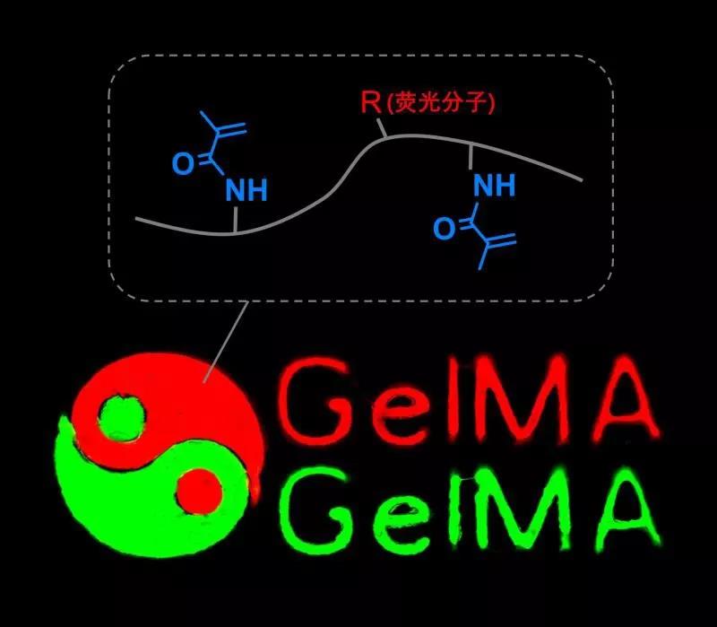
The fluorescent GelMA developed by the team is achieved by "grafting" fluorescent molecules on GelMA, which has a specific fluorescent color due to different grafted fluorescent molecules. This chemical labeling method avoids the shortcomings that fluorescent molecules easily diffuse out of the system in methods such as physical mixing or electrostatic adsorption, and it also avoids the shortcomings of uneven imaging of fluorescent particles.

2. Advantages of fluorescent GelMA
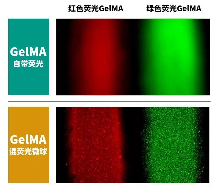
1. Fluorescent GelMA imaging is uniform. The following figure shows the imaging comparison of two GelMA with self-fluorescent and mixed fluorescent microspheres. The fluorescent GelMA imaging is uniform and continuous, and the surface of the mixed fluorescent microspheres GelMA has an obvious graininess.

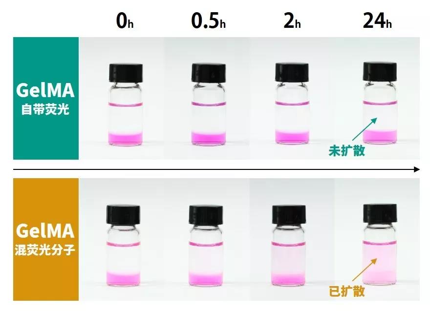
2. Fluorescent GelMA is stable and does not diffuse. The figure below shows the diffusion comparison of two kinds of GelMA with self-fluorescent and mixed fluorescent molecules. GelMA mixed with fluorescent molecules is easy to diffuse; self-fluorescent GelMA can maintain fluorescence characteristics for a long time.

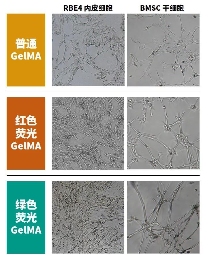
3. Fluorescent GelMA has excellent biocompatibility. The figure below shows the growth status of endothelial cells (RBE4) and bone stem cells (BMSC) cultured on the fifth day using fluorescent GelMA and ordinary GelMA. Fluorescent GelMA has the same excellent biocompatibility as ordinary GelMA.

Three, fluorescent GelMA application case
1.3D printing. In recent years, GelMA has become the star ink of 3D bioprinting. Fluorescent GelMA can make the characterization of the printed structure more clear and stable. The fluorescent GelMA developed by the team can be applied to all types of biological 3D printers. The picture below shows the fluorescent cool structure made by the BKNM-BP series of biological 3D printers.

2. Fluorescence tracer. Using fluorescent GelMA can easily track the changes of GelMA or other carriers in the body. Self-fluorescent GelMA will not diffuse in the body and can truly track the distribution of fluorescent substances.

| Warm tips: Suzhou Beike nano products are only used for scientific research, not for human body,different batches of products have different specifications and performance |
Message
|
|
 |
Scan code concerns WeChat official account
QQCommunication group:1092348845
|
|
| Warm tips: Suzhou Beike nano products are only used for scientific research, not for human body,different batches of products have different specifications and performance.The website pictures are from the Internet. The pictures are for reference only. Please take the real object as the standard. In case of infringement, please contact us to delete them immediately. |